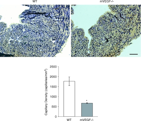
Figure 4. Alkaline phosphatase staining of cardiac muscle sections and average capillary density for wild-type (WT) control and muscle VEGF-deficient (mVEGF−/−) mice.
A, representative images showing alkaline phosphatase stained cardiac muscle sections from WT and mVEGF−/− mice. Images shown at 12.5× magnification (Bar = 5 μm). Capillaries appear as dark purple stained. B, average capillary density (number of capillary per mm2) in WT and VEGF deficient (mVEGF−/−) mice (n= 3/group). Data are means ±s.e.m. *Significantly different compared to WT (P < 0.05).

