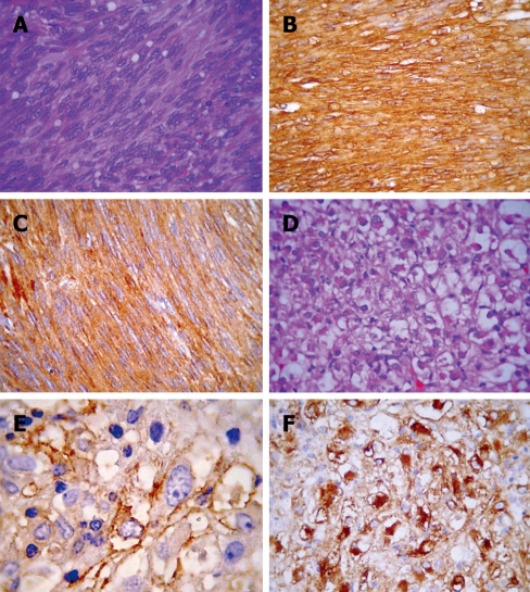
Figure 2.
Morphological and immunohistochemical features of gastric GIST (A-C) and hepatic PEComa (D-F). A: GIST with characteristic spindled bipolar cells with occasional paranuclear cytoplasmic vacuoles; B and C: Strong staining for CD117 and CD34, respectively; D: Hepatic PEComa with a monotonous epithelioid morphology with cytoplasmic clearing that is hardly distinguishable from epithelioid GIST (note scattered cells with rhabdoid cytoplasm); E: A coarsely granular HMB45 reactivity; F: HH35 showing a strong paranuclear cytoplasmic reactivity.

