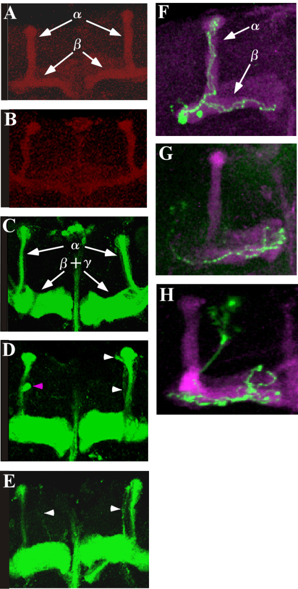
Figure 6.

Morphology of alpha/beta lobes and alpha/beta neurons in mushroom bodies of the wild-type and the dmPsGEFΔ21 mutant. The alpha lobes detected by Fas II staining are thinner in the dmPsGEFΔ21 mutants (B) than in the wild-type (A). The alpha and beta lobes are indicated by arrows in (A). The alpha lobes of late-born alpha/beta neurons are visualized by 201Y-Gal4 and UAS-mCD8::GFP in the wild-type (C) and dmPsGEFΔ21 mutants (D and E). The alpha lobes are thinner in dmPsGEFΔ21 mutant than in the wild-type, as observed above. In addition, the alpha lobes are often short (magenta arrowhead in D), and their positions along the beta lobes vary (white arrowheads in D and E). The alpha as well as the beta and gamma lobes are indicated by arrows in (C). Findings of the repressible cell marker analysis reveal that the axons of wild-type alpha/beta neurons (green) bifurcated toward the alpha/beta lobes (magenta) (F). However, the axons of dmPsGEFΔ21 alpha/beta neurons (green) often fail to grow toward the alpha lobes (G and H). The alpha and beta lobes are indicated by arrows in (F).
