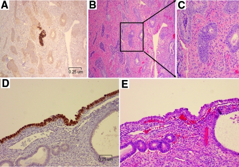
Figure 2.
p53 signatures and their corresponding H&E endometrial glands. Lower panel showed representative endometrial glands from case 2 with p53 signatures (A, glandular pattern), the corresponding H&E section (B), and the morphologically magnified p53 signature gland (C). Lower panel showed pictures from case 9 with a surface pattern of p53 signature (D, surface pattern) and the corresponding area of H&E section (E). Morphologically, nuclear atypia of the epithelia with p53 signatures was inconspicuous. (Magnification of A, B, D, and E = original ×200).

