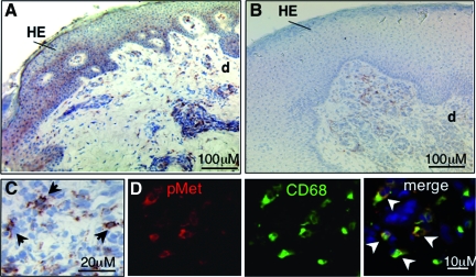
Figure 9.
c-Met phosphorylation in wound repair. Immunohistological analysis of cryosections from healing (A, C, D, day 8 after wounding) and non-healing (B) wounds using an anti-phospho-c-Met antibody; brown staining indicates phospho-c-Met positive cells. Note that healing wounds show phosphorylation of c-Met in the hyperproliferative epidermal (HE) wound edge and papillary dermis, whereas non-healing wounds show staining exclusively in dermal cells. Arrows in (C) indicate cells positive for c-Met phosphorylation. D: Predominantly macrophages (CD68) (indicated by arrowheads) display phosphorylation of c-Met. e, epidermis; d, dermis.

