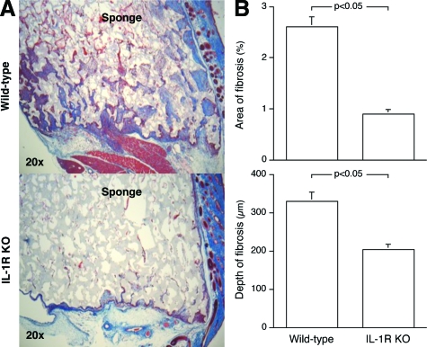
Figure 1.
IL-1 signaling is essential for the fibrotic response seen in deep tissue sterile wounds. PVA sponges were implanted into IL-1R KO or wild-type mice and harvested 14 days later. A: Low-power photomicrographs of trichrome-stained sponge wounds. Sponge material stains lightly, whereas fibrotic tissue appears dark blue. Sirius red staining confirmed that the areas of fibrosis stained by trichrome reflect collagen deposition (not shown). Overlying skin is oriented to the right. B: Quantitative histomorphometric analysis of the sponge wounds, expressed as total area of fibrosis per microscopic field and depth of fibrotic buds. n = 6 animals per group, Mann Whitney’s U-test (P < 0.05).

