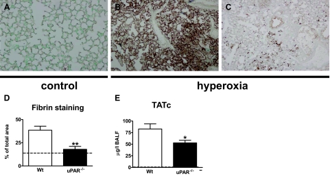
Figure 7.
uPAR−/− mice demonstrate decreased local activation of coagulation. Wild-type and uPAR−/− mice were exposed to 80% oxygen. Control mice were exposed to room air. Representative fibrin(ogen) immunostaining of lung tissue of control mice (A) and of wild-type and uPAR−/− mice exposed to hyperoxia for 4 days (B and C, respectively). Original magnification ×4. Graphical representation of the % of the total area with positive fibrin(ogen) staining (D) according to the scoring system described in the Materials and Methods section. Thrombin-antithrombin complex (TATc) concentrations were measured in BALF (E). Data are mean ± SE of 7 to 10 mice per time point. *P < 0.05 versus wild-type mice; **P < 0.01 versus wild-type mice. Dotted lines represent the mean values obtained from normal mice (exposed to room air).

