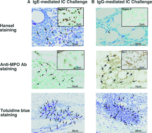
Figure 4.
Histological tissue sections showing eosinophil, neutrophil and mast cell accumulation of wild-type mice at 24 hours after IC challenge by intradermal injection of IgE (A) or (B) IgG anti-TNP. Hansel-staining (anti-Siglec-F Ab; insets) shows eosinophils (arrows; upper panels), and anti-MPO Ab staining (anti-Gr-1 Ab: insets) demonstrates neutrophils (arrows; middle panels). Mast cells were revealed by toluidine blue-staining (arrows; bottom panels). Original magnification = ×100 (toluidine blue) and ×200 (Hansel, anti-Siglec-F, anti-MPO, and anti-Gr-1).

