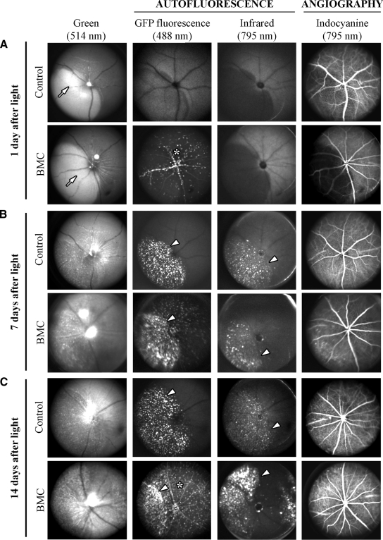
Figure 1.
SLO and angiography in BMC and wild-type mice. A: One day after light exposure, wild-type (control) and BMC mice showed a subretinal edema in the hot spot (arrow), which appeared lighter than the remaining fundus in the 514 nm-funduscopy. Fluorescence of bone marrow-derived cells was distinct within vessels and in retinal tissue (asterisk) of BMC mice. Indocyanine green angiography showed no leakage of retinal and choroidal vessels in control and BMC mice. B: Seven days after light exposure, in addition to GFP fluorescence of cells in BMC, both control and BMC mice displayed distinct autofluorescent deposits in the hot spot absorbing at 488 and 795 nm (arrowheads). C: Fourteen days after light exposure, accumulation of retinoid-containing cellular debris in the hot spot region persisted (arrowheads) and GFP fluorescent cells were distributed in the entire retina (asterisk).

