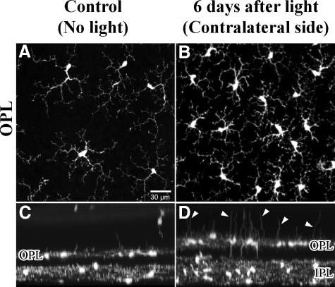
Figure 5.
Confocal light micrographs of CX3CR1−/− control retinas and 6 days after light exposure. A and C: Controls. A: View onto flat mount of the OPL. C: The same retinal area as shown in A, but tilted by 90°. In control retinas, microglial cells of the OPL showed characteristic arborizations of their processes and only few processes extending toward the photoreceptor layer (outer nuclear layer). B and D: In the contralateral retinal area from the light-exposed eye, a significant increase in number of microglial cells occurred 6 days after light exposure with a remarkable increase of thickened processes extending toward the photoreceptor layer (arrowheads in D).

