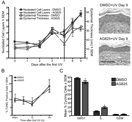
Figure 2.
Inhibition of Erbb2 suppresses UV-induced hyperplasia and proliferation. Groups of AG825-and DMSO-pretreated mice were exposed to 10 kJ/m2 UV twice with an interval of 1 week, as described in Figure 1A. Mice were euthanized at the indicated time points following irradiation. A: Quantification of epidermal hyperplasia from at least three mice per group was averaged and the mean graphed. Vertical dashed lines indicate UV exposure. Representative H&E sections shown on right. Scale bar = 100 μm. B: The percent basal cells positive for terminal deoxynucleotidyl transferase-mediated dUTP nick end labeling was quantified in skin sections at the indicated time points following the second UV irradiation. N ≥3. C: DNA content flow cytometry was performed using sections of skin from mice euthanized 2 days after the second UV exposure and the percentage of cycling cells graphed. N ≥3 mice. Treatment is significantly different using a Bonferroni posttest, where *P ≤ 0.05.

