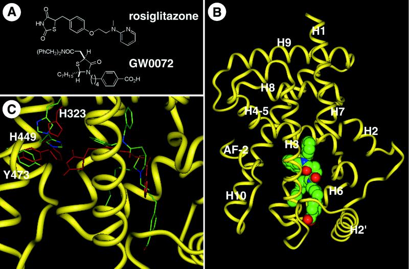
Figure 1.
GW0072 is a PPARγ ligand with a unique binding mode. (A) Chemical structures of the TZD rosiglitazone and the thiazolidine acetamide GW0072. (B) Cocrystal structure of the PPARγ ligand-binding domain with GW0072. The polypeptide backbone is illustrated as a yellow worm schematic, and GW0072 is shown as a Van der Waals space-filling representation with each atom type colored: carbon, green; oxygen, red; nitrogen, blue; and sulfur, yellow. (C) The PPARγ polypeptide backbone atoms from the cocrystal structure with GW0072 superimposed on the cocrystal structure with rosiglitazone (6). The polypeptide backbone of the GW0072 cocrystal structure is shown as a thin yellow ribbon; GW0072 as well as H323, H449, and Y473 of the protein complex are shown as carbon, green; oxygen, red; nitrogen, blue; and sulfur, yellow. Rosiglitazone and the same three amino acids for its respective protein complex are colored red. Differences between the rosiglitazone and GW0072 cocrystal structures exist in the side-chain amino acid positions and not in the overall polypeptide conformation except for the loop between helix 2′ and helix 3, which is the result of crystal packing. GW0072 occupies an epitope of the ligand-binding pocket of PPARγ that precludes it from interactions with Y473, H323, and H449.

