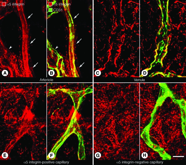
Figure 1.
Distribution of α5 integrin immunoreactivity in normal tracheal blood vessels. Confocal microscopic images of tracheal whole mounts stained for CD31 (green, blood vessels) and α5 integrin (red) immunoreactivities of a small arteriole (A and B, arrows), venule (C and D), and unusual capillary (E and F) from a pathogen-free mouse. G and H: Tracheal capillary, like most tracheal capillaries, without detectable α5 integrin immunoreactivity. Arrowheads in A and B mark a small venule. Scale bar = 10 μm.

