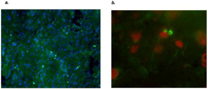
Figure 4.
There are multiple perforin positive cells (stained with FITC) within the infarcted tissue of this LPS treated animals sacrificed one month after MCAO (a). These perforin positive cells are seen in close juxtaposition to DAPI positive cells (40x). The DAPI positive cells were shown to be neurons using a Texas-red conjugated antibody for neuron specific enolase (100x) (b).

