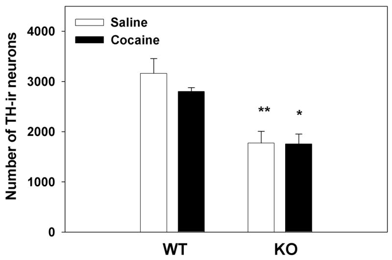
Figure 1. Stereological analysis of TH expression in the VTA of WT and nNOS KO mice after saline and cocaine administration.

WT and nNOS KO mice received saline or cocaine (20mg/kg) for 5 consecutive days. After 24 hr animals were perfused and brain tissue was prepared for staining of TH-ir neurons, as described in Materials and Methods. A significant genotype-dependent effect was observed (**p<0.01 and *p<0.05) as the number of TH-ir neurons in the VTA of nNOS KO mice was lower than that in their WT counterparts, regardless of cocaine treatment which had no significant effect on the number of VTA TH-ir neurons.
