
Figure 4. WNV infection induces expression of selective cell adhesion molecules in HBMVE cells.
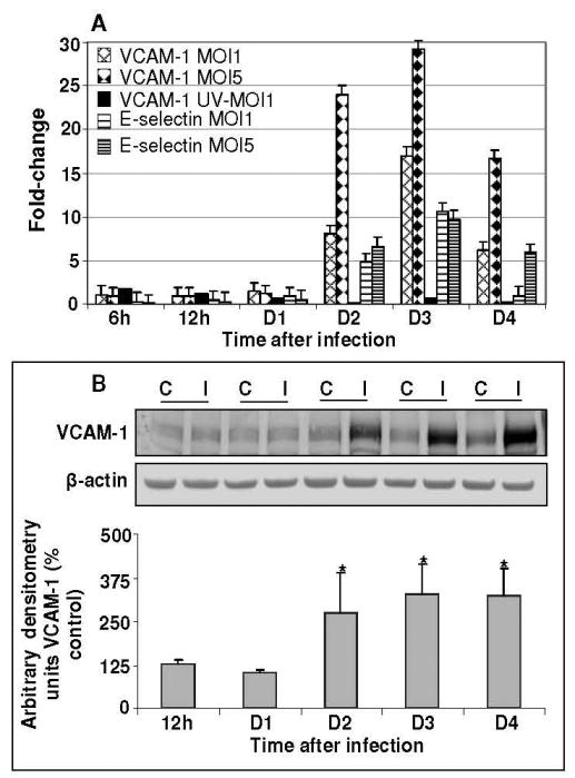
(A) cDNA templates from mock, UV-WNV and WNV-infected HBMVE cells at different time points after infection were used to determine the fold-change of VCAM-1 and E-selectin as compared to mock-infected controls at same time point. Data represents mean of four independent experiments conducted in duplicate. (B) 50-70 μg of cellular proteins extracted from mock- and WNV-infected HBMVE cells were separated on PAGE, transferred onto PVDF membranes and immunoblotted with antibodies specific to VCAM-1 and β-actin. Blots were scanned using BioRad Phosphorimager and analyzed using Quantity one program. Data is represented as percentage of un-infected HBMVE cells at the corresponding time point. Mean comparisons were based upon extrapolated CI for three data points. *p<0.05, compared to corresponding mock-infected cells. C, mock and I, WNV infected.