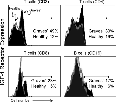
Figure 1.
Increased frequency of IGF-IR-expressing T and B lymphocytes from monozygotic twins with GD. Expression of IGF-IR by CD3+ T cells (upper left panel), CD4+ T cell subset (upper right panel), CD8+ T cell subset (lower left panel), and CD19+ B cells (lower right panel) from twins with GD (solid black histograms) and those not manifesting the disease (open gray histograms). Representative histograms (GD, n = 15; healthy, n = 21).

