
Figure 4. Sar1-GFP accumulates in and around the actin-coated structures in Bic- C mutants.
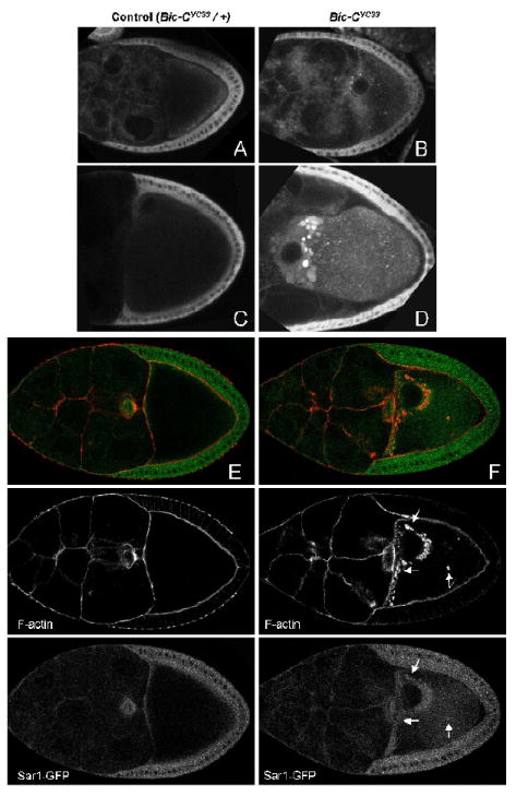
Live imaged (A-D) and fixed (E, F) egg chambers expressing Sar1-GFP in a heterozygous (A, C, E) or homozygous Bic-CYC33 mutant (B, D, F) background. In live imaged homozygous Bic-C mutant oocytes, luminous foci of Sar1-GFP accumulate at the anterior of the oocyte. In fixed samples, these foci clearly overlap with the actin-coated structures in Bic-C (F, see arrows).