
Figure 1. Photomicrographs of nitrotyrosine immunohistochemistry.
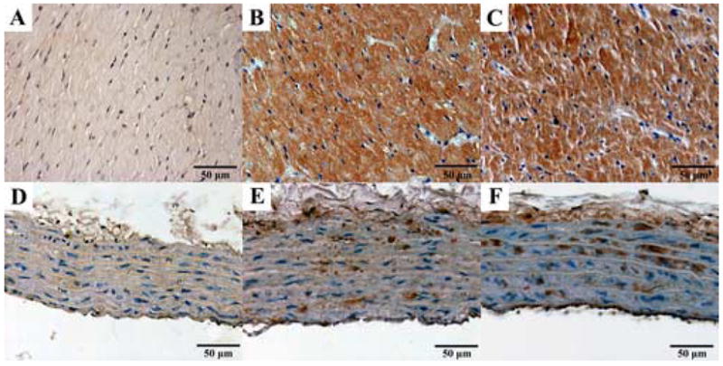
Representative immunohistochemical stainings for nitrotyrosine (NT, brown staining) in the myocardium (A–C) and aortic wall (D–F). Young control group: A, D; aging control group: B, E; and aging INO-1001 treatment groups: C, F (magnification: 400X, scale bar: 50μm).