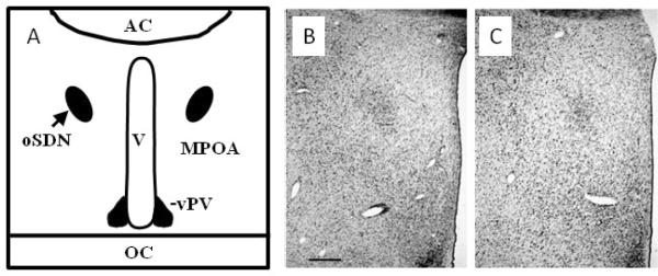
Figure 2.
A)Schematic diagram of a coronal section through the sheep at the level of the optic chiasm (OC) and anterior commissure (AC). The position of the oSDN is shown bilateral to the third ventricle (V) in the central part of the medial preoptic nucleus (MPOA). The ventral paraventricular nucleus (vPV) is shown at the base of the ventricle. B) Micrograph of oSDN from the left hypothalamus of a female-oriented ram. The third ventricle is at the right of the figure. C) Section of a male-oriented ram comparable to that in (B). The photomicrograph are taken near the midpoint of the anterior - posterior extent of the oSDN. The scale bar is 1 mm.

