
Figure 6.
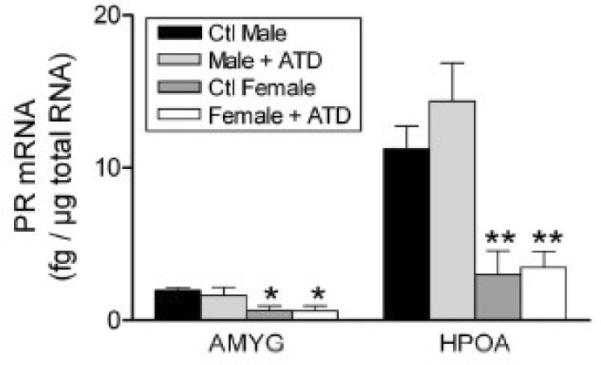
Sex differences in the expression of progesterone receptor mRNA in the amygdala (AMYG) and hypothalamus-preoptic area (HPOA) of gestational day 64 fetal lambs. The levels of progesterone receptor mRNA were measured using a RNase protection assay that measured mRNA concentration using a standard curve of progesterone receptor sense RNA and normalized values to levels of cyclophilin mRNA in each sample. Data are presented as means ± SEM, n =3- 4/group. *, P < 0.5; **, P < 01 male versus female. Some fetuses were exposed to the aromatase ATD given by Silastic implants to their mothers as described in (Roselli, Resko, and Stormshak, 2003). This treatment had no effect on progesterone receptor mRNA expression. Reprinted from Roselli et al. (Roselli et al., 2006).