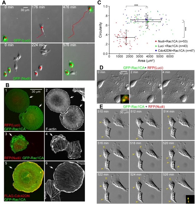
Figure 1. Nudel knockdown impairs cell spreading.
(A) Nudel RNAi inhibits lamellipodial formation and cell migration. Representative image sequences are shown for ECV304 cells transfected with pTER-Nudi-GFP or pTER-Luci-GFP (green) for approximately 60 h. Red lines indicate cell tracks. See also Videos S1 and S2. (B and C) Nudel RNAi represses Rac1CA-stimulated cell spreading independently of Cdc42 activity. Panels 1–4: ECV304 cells were transfected for approximately 60 h with an indicated RNAi construct and then transfected again to express GFP-Rac1CA for approximately 12 h. Panels 5 and 6: cells were cotransfected for approximately 12 h to coexpress FLAG-Cdc42DN and GFP-Rac1CA. Arrows indicate transfectants. The area and circularity (4π×area/perimeter2) are used to reflect the extent of cell spreading. Error bars show SD. Asterisks indicate p<0.005. (D and E) Time-lapse images of typical control or Nudel-depleted cells overexpressing GFP-Rac1CA. Membrane protrusions (arrowheads) fail to be stabilized upon Nudel RNAi. Transfectants were identified through their coexpression of both RFP and GFP (insets). See also Videos S3 and S4.

