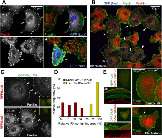
Figure 2. Effects of Nudel knockdown on FCs and stress fibers.
(A and B) Nudel RNAi results in large peripheral FAs associated with thick stress fibers due to the collapse of cell edges (A). ECV304 cells were transfected with an indicated RNAi plasmid for approximately 72 h (arrows). Arrowheads indicate lamellipodia. In (B), cells were treated with 20 µM blebbistatin for 45 min to abolish myosin II-mediated tensions on stress fibers. More stress fibers and FAs were preserved in Nudel-depleted cells (arrows) after the drug treatment. As a result, untransfected cells generally contained higher levels of free paxillin and thus exhibited stronger cytosolic paxillin staining. (C and D) Nudel depletion attenuates FX formation even in the presence of GFP-Rac1CA. ECV304 cells were transfected as described in Figure 1B. Arrowheads indicate typical FXs. Relative FX-containing area was calculated as the percentage of radians of the FX-containing sector in a circle, centered at the centroid of a cell using ImageJ. (E) Nudel is important for nascent adhesion formation. ECV304 cells were transfected as described in Figure 1B and treated with 20 µM blebbistatin for 25 min prior to fixation to block the transition of nascent adhesions to FXs [5]. For four-color staining, F-actin was decorated with phalloidin-TRITC, whereas secondary antibodies conjugated with Alexa-405 and 647 were used with rabbit anti-FLAG antibody and anti-paxillin mAb to label FLAG-Rac1CA and paxillin. GFP was visualized through its autofluorescence. A typical region (arrow) was magnified to show details.

