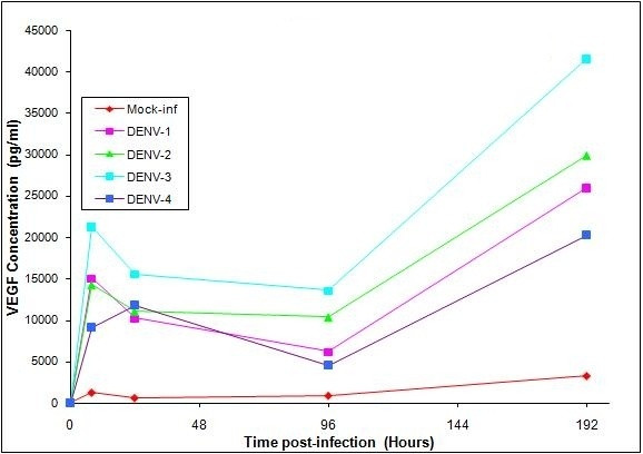
Figure 2.

VEGF Production in HPMEC-ST1.6R Cells infected with DENV-1, -2, -3, and -4. Monolayers of HPMEC-ST1.6R cells were infected with either DENV-1, -2, -3 or DENV-4 viruses as previously described [6], and incubated at 37°C in 5% CO2 for 6 days. The cell media of DENV-infected cells and mock-infected cells were collected at 0, 8, 24, 96 and 192 hours post-infection, and analyzed for VEGF production [actual values listed in Table 1]. Mean VEGF levels were determined using a VEGF analyte detection kit and a BioPlex suspension array analyzer from BioRad. Significant increases (p ≤ 0.05) in cytokines between virus-infected and mock-infected cell cultures are given in Table 2. [Bars equal standard deviation from the mean of triplicates].
