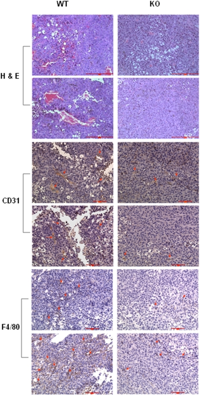
Figure 11. Immunohistochemistry staining of melanoma tissue from WT and KO mice.
H & E staining of tumor sections for gross observation. Staining of tumor section with anti-CD31 (1∶50) for angiogenesis analysis. Staining of tumor section with anti-F4/80 (1∶75) to view macrophage infiltration. Representative positively stained cells were indicated by arrows. Scale bar = 100 µm.

