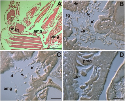
Figure 7. TUNELPOS promastigotes are present in the intestinal tract of infected sand flies.
Lutzomyia longipalpis sand flies were fed with mice blood containing infected macrophages. After 10 days female insects were fixed and histochemical analysis for TUNEL labeling was performed. (A) Hematoxilin-eosin stain showing overview of sand fly section. (B) Parasite concentration at the bulbous cardia region of the foregut and isolated and clumped TUNELPOS promastigotes in the foregut (fg). Most of the stained parasites are already acquiring a round-shaped morphology (arrows). (C) and (D) elongated TUNELNEG promastigotes in respectively the anterior midgut (amg) and posterior midgut (pmg) of infected sand flies (arrowheads). Bars represent 200 µm (A) and 20 µm (B, C and D). Asterisks on panels A and B indicate the bulbous cardia region of the foregut.

