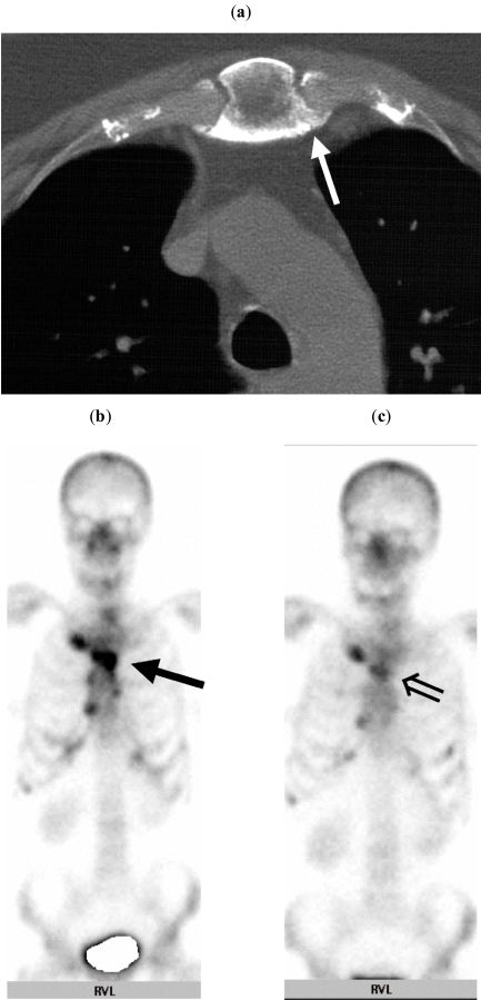
Fig (2).
(a) Axial CT scan of the sternal region reveals marked hyperostosis and osteitis of the sternum (↑). (b) Technetium-99m bone scintigram shows considerable activity in the sternoclavicular, sternocostal joints and the vertebral column (↑). (c) After 26 months of therapy, overall signal intensities have decreased substantially in the sternal region (⇑).

