Fig. 1.
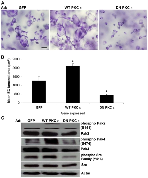
PKCε stimulates EC lumen formation in 3D collagen matrices. (A) ECs infected with adenoviruses (Ad) expressing GFP, WT-PKCε or DN-PKCε were suspended within 3D collagen matrices for 24 hours. Scale bar: 50 μm. (B) Quantification of EC lumen formation at 24 hours. Data are shown as mean EC lumenal area ± s.d. (n=3). *P<0.05 compared with GFP control. (C) EC extracts were prepared at 24 hours for western blot analysis and probed for phospho-Pak2, phospho-Pak4, phospho-Src or Pak2, Pak4, Src and actin controls. The actin control blot was derived from cut lanes of the same gel and a single exposure.

