Fig. 7.
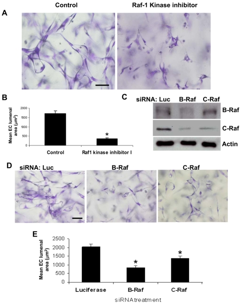
Inhibition of Raf kinases blocks EC lumen formation in 3D collagen matrices. (A,B) ECs were resuspended in 3D collagen matrices for 24 hours in the absence or presence of Raf1 kinase inhibitor GW5074 (5 μM). (A) Representative fields of EC lumen formation assay. Scale bar: 50 μm. (B) Quantification of EC lumen formation. (C-E) siRNA suppression of B-Raf or C-Raf inhibits EC lumen formation in 3D collagen matrices. (C) Lysates were prepared for western blot analysis and probed for phospho-B-Raf, phospho-C-Raf and actin control. ECs treated with the indicated siRNAs were resuspended in 3D collagen matrices for 24 hours. (D) Representative fields of EC-lumen-formation assay. Scale bar: 50 μm. (E) Quantification of EC-lumen-formation assay. Data are shown as the mean EC lumenal area ± s.d. (n=3). *P<0.01 compared with control.

