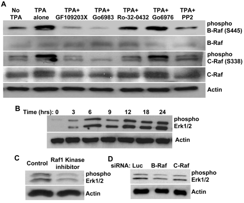
Fig. 9.
B-Raf and C-Raf activation occur downstream of PKCε and SFKs during EC-lumen-formation events in 3D collagen matrices. (A) ECs were resuspended in 3D collagen matrices in the absence or presence of TPA, GF109203X (2.5 μM), Ro-32-0432 (5 μM), Go6983 (5 μM), Go6976 (5 μM) or PP2 (10 μM). Lysates were prepared at 24 hours for western blot analysis and probed for phospho-B-Raf, phospho-C-Raf, B-Raf, C-Raf or actin. (B-D) ERK1/2 proteins are phosphorylated during EC lumen formation in 3D collagen matrices. (B) Extracts of EC cultures were prepared at the indicated time points and probed for phospho-ERK1/2 or actin. (C) ECs were resuspended in 3D collagen matrices for 24 hours in the absence or presence of Raf-kinase inhibitor, GW5074 (5 μM). Lysates were prepared for western blot analysis and probed for phospho-ERK1/2 or actin. (D) ECs treated with the indicated siRNAs were resuspended in 3D collagen matrices. Lysates were prepared for western blot analysis and probed for phospho-ERK1/2 or actin.

