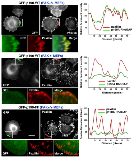
Fig. 4.
FAK and p190A tyrosine phosphorylation promote p190A FA localization upon FN plating. FAK+/+ or FAK–/– MEFs were transfected with GFP–p190-WT or GFP–p190-FF, serum starved, plated onto FN (10 μg/ml, 30 minutes), and analyzed for GFP and paxillin colocalization. Fluorescence-intensity profiles represent the area marked by the colored lines (green p190A, red paxillin). Insets, enlargement of the area of interest shown for GFP-p190A, paxillin and the merged images. GFP–p190-WT and paxillin show colocalization at peripheral FAs in FAK+/+ but not in FAK–/– MEFs as determined by fluorescent-intensity overlap. The GFP–p190-FF fluorescent-intensity profile does not overlap with paxillin at FAs. Scale bar: 20 μm.

