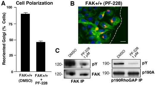
Fig. 5.
Pharmacological FAK inhibition blocks MEF polarization and p190A tyrosine phosphorylation. (A) Golgi-reorientation analyses of scratch-wounded FAK+/+ MEFs performed in the presence of DMSO or 1 μM PF-228 (FAK inhibitor). Data is the percentage of 100 cells with Golgi that were reoriented towards the wound edge (± s.d.). (B) Representative image from a wound healing assay. MEFs were stained with β-Cop antibody (red), anti-tubulin antibody (green) and Hoechst nuclear dye (blue). The position of leading lamella (broken white line) is shown. Scale bar: 30 μm. (C) FAK and p190A IPs from FN-plated MEFs (30 minutes) in the presence of DMSO or PF-228 (1 μM) were sequentially analyzed by anti-pY followed by anti-FAK or anti-p190A immunoblotting, respectively.

