Fig. 1.
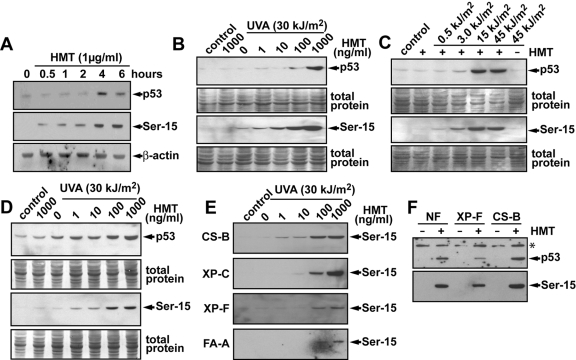
Induction of Ser-15 phosphorylation and p53 protein levels by PUVA. A, human diploid fibroblasts were treated with HMT (1 μg/ml = 3.8 μM) and irradiated with 30 kJ/m2 with UVA (10 min). Cells were incubated in fresh media at 37°C and harvested at different times, and the levels of total p53 protein and Ser-15 phosphorylated p53 were determined by Western blotting. β-Actin was used as a loading control. B, human diploid fibroblasts were treated with different doses of HMT followed by UVA irradiation (30 kJ/m2). Cells were collected 6 h later, and the levels of total p53 protein and Ser-15 phosphorylated p53 were determined by Western blotting. After exposure to film, the membranes were stained with Coomassie blue stain to visualize total protein loading and transfer in the different lanes. C, human diploid fibroblasts were treated with HMT (1 μg/ml = 3.8 μM) and irradiated with different doses of UVA. Cells were harvested 6 h later, and the levels of total p53 protein and Ser-15 phosphorylated p53 were determined by Western blotting. Note that neither HMT nor UVA (45 kJ/m2) alone induced p53 or phosphorylation of p53, whereas HMT with the higher exposure of UVA light induced high levels of both. D, diploid fibroblasts derived from a patient with xeroderma pigmentosum complementation group A (XP-A) were treated as in B. E, diploid fibroblasts from various DNA repair-deficiency syndromes were treated as described in B and analyzed for Ser-15 phosphorylation. F, diploid normal, XP-F, and CS-B fibroblasts were treated with 100 ng/ml (380 nM) HMT and 30 kJ/m2 UVA and incubated for 24 h before the cellular levels of p53 and Ser-15 phosphorylated p53 were determined by Western blot. *, a nonspecific band was used to assess loading.

