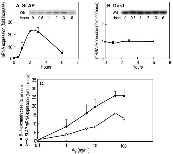
Fig. 1.
Increased expression of SLAP mRNA and protein in response to Ag-stimulation. RBL-2H3 cells sensitized overnight with 50 ng/ml DNP-specifc IgE were stimulated with 50 ng/ml Ag. Panels A and B, cells were stimulated for the indicated times and relative levels of DOK1 and SLAP mRNA and protein (insets) were determined by real-time PCR and immunoblotting respectively. Error bars show Standard Deviations for PCR determinations. The data indicate relative levels of mRNA (zero hours equals 1) and are from one of two similar experiments. Panel C compares the extent of increase of SLAP protein and degranulation with different concentrations of Ag. SLAP was measured 3 h and degranulation 20 min after addition of Ag. Degranulation was assessed by calculating per cent of cellular β-hexosaminidase (a granule marker) that was released into the medium. Data (mean ± SEM) were from four separate cultures and is representative of two experiments.

