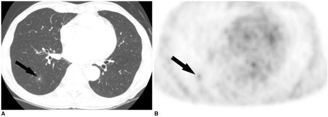
Fig. 3.
Simple pulmonary eosinophilia in a 65-year-old man with esophageal cancer that mimicked metastasis on both CT and PET.
A. The transverse CT scan obtained with the lung window setting shows a ground-glass opacity nodule with a central solid portion in the right lower lobe (arrow).
B. The tansverse FDG PET scan shows the increased uptake in the nodule with SUV of 2.0 (arrow).

