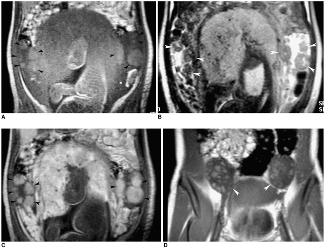
Fig. 1.
Pregnancy luteoma. The peripheral-located multinodular ovarian masses (arrowheads) show intermediate high signal intensity on the coronal T1-weighted image (A, 101/4/1 [repetition time/echo time/excitation]), low signal intensity on the T2-weighted image (B, 4/90/1), and avid enhancement on the Gd-DTPA enhanced T1-weighted image (C, 101/4/1). Three weeks postpartum, the coronal Gd-DTPA enhanced T1-weighted image (D) show the greatly diminished size of the enhancing ovarian masses, although the ovaries (arrowheads) remain large.

