Fig. 3.
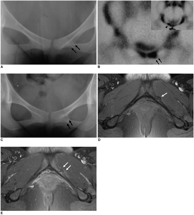
A 19-year-old female military cadet with a fatigue fracture of the left inferior pubic ramus.
A. Initial radiograph only shows indistinct fracture line in the left inferior pubic ramus (arrows).
B. Bone scan with 99mTc-MDP reveals a focal area of increased tracer activity in the left inferior pubic ramus (arrows), suggesting a fatigue fracture, which is disclosed definitely on the caudal view of the pelvis (arrowhead).
C. Two months later, the follow-up plain radiograph reveals discrete callus formation in the left inferior pubic ramus (arrows).
D. Axial T1-weighted MR image demonstrates a fracture line and a minimally displaced small bony fragment with callus formation in the anterior portion of the left inferior pubic ramus (arrow), suggesting an avulsion type of fatigue fracture.
E. Gadolinium-enhanced MR image shows soft tissue enhancement around the callus formation (arrows).

