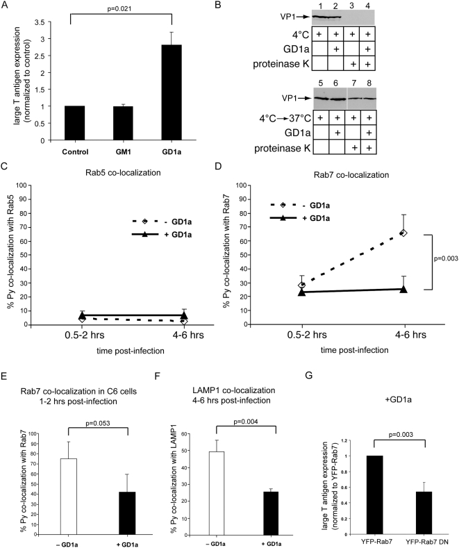
Figure 4. Decreased co-localization of Py with the late endosome and lysosome in GD1a-supplemented cells.
(A) NIH 3T3 cells were incubated with purified GM1 or GD1a, washed, infected with Py, and the extent of infection was assessed as in Figure 1B. Results were normalized to non-supplemented cells (control cells). In the control cells, 43/984 cells expressed large T antigen. (B) Untreated (control) or GD1a-supplemented NIH 3T3 cells were incubated with Py at 4°C to allow viral binding and then treated with proteinase K where indicated (top panel) or incubated at 37°C for 1 hr before proteinase K treatment to determine viral entry (bottom panel). (C, D) The extent of co-localization of labeled Py with (C) Rab5-containing vesicles or (D) Rab7-containing vesicles at the early (0.5–2 hrs) and late (4–6 hrs) time points in both control and GD1a-supplemented NIH 3T3 cells. (E) Co-localization of labeled Py with Rab7-containing vesicles at 1–2 hrs post-infection in the ganglioside-deficient C6 cells. (F) Co-localization of labeled Py with LAMP1-containing vesicles 4–6 hrs post-infection in NIH 3T3 cells. (G) The extent of Py infection in GD1a-supplemented cells expressing wild type YFP-Rab7 or dominant negative YFP-Rab7 (DN). At least 220 transfected cells were analyzed from three independent experiments. All data are the mean+/−SD. A two-tailed t test was used.

