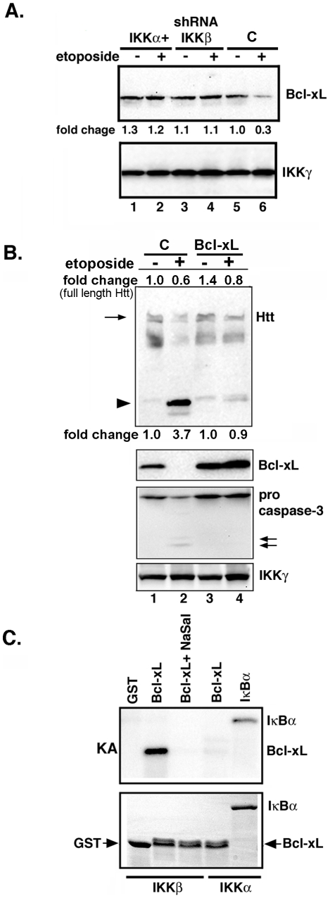
Figure 6. Etoposide promotes reduction of Bcl-xL.
(A) Extracts of control and etoposide-treated MESC2.10 neurons were examined for Bcl-xL by Western blotting. Neurons were treated with etoposide for 6 hrs. Top panel shows staining for Bcl-xL and the bottom panel indicates IKKγ as a loading control. Fold changes were normalized to the intensity of loading control, and compared to that of untreated control neurons (lane 5). (B) Bcl-xL expression prevents Htt proteolysis by etoposide. MESC2.10 neuroblasts were transduced with a lentivirus expressing Bcl-xL (Lanes 3 and 4) and treated with etoposide for 6 hrs. EGFP-lentivirus was used a control (C). Top panel Western shows blotting for Htt. Arrow indicates the full-length Htt and the arrowhead shows the cleaved Htt products. Second and third panels show staining for Bcl-xL and pro-caspase-3, respectively. IKKγ levels were used as a loading control. (C) IKKβ phosphorylates Bcl-xL. Active recombinant IKKα or IKKβ were tested for the ability to phosphorylate Bcl-xL. The kinase assay was performed as described in the M&M with recombinant Bcl-xL as a substrate. Products were visualized by autoradiography. IκBα was used as a positive control substrate for IKKα. The top panel shows the kinase product (KA) and bottom panel shows the SDS-PAGE and coomassie-blue staining of the substrates use in kinase assays.

