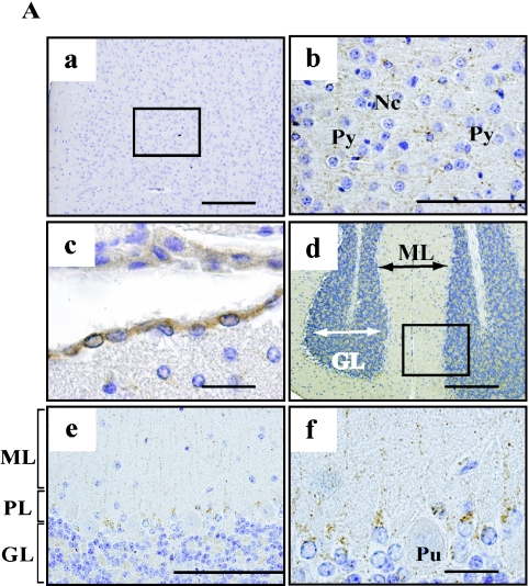
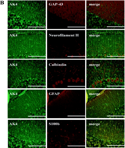
Fig. 2.
Cellular localization of AK4 in the brain. (A) Immunohistochemical analyses were performed using anti-AK4. a, b; Cerebral cortex, c; Choroid plexus, d, e, f; Cerebellum. Bar=200 µm (a, d), 100 µm (b, e), and 20 µm (c, f). Py, pyramidal cell; Nc, neuroglial cell; GL, granular layer; ML, molecular layer; PL, Purkinje layer; Pu, Purkinje cell. Open square indicates the area of higher magnification. (B) Double immunodetection with anti-AK4 and neural or glial marker antibodies. Bar=100 µm.


