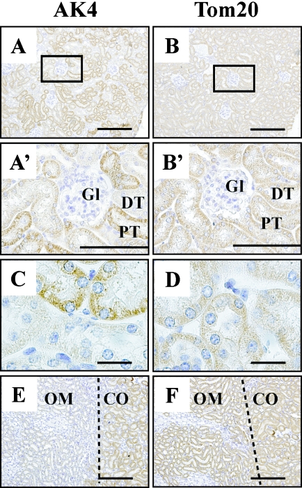
Fig. 4.
Cellular localization of AK4 in the kidney. (A, A’, B, B’, C, D) Renal cortex. (E, F) Outer layer of renal medulla. Immunohistochemical analyses were performed using anti-AK4 (A, A’, C, E) and anti-Tom20 antibodies (B, B’, D, F). Bar=200 µm (A, B, E, F), 100 µm (A’, B’), and 20 µm (C, D). Gl, glomerulus; DT, distal tubule; PT, proximal tubule; OM, outer zone of medulla; CO, cortex. Open square indicates area of higher magnification. Dotted line in C, D shows the boundary between the outer zone of the medulla and the cortex.

