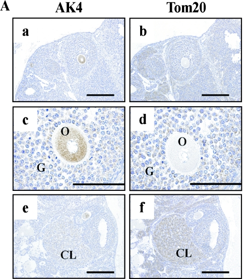
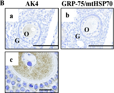
Fig. 8.
Cellular localization of AK4 in the ovary. Immunohistochemical analyses were performed using anti-AK4 (A-a, c, e, B-a, c), anti-Tom20 (A-b, d, f), and anti-GRP-75/mtHSP70 (B-b). Overviews at low magnification (A-a, b, e, f) and high magnification (A-c, d, B-a, b, c). (A-a–d, B-a, b, c) Oocytes and granulose cells. (A-e, f) Corpus luteum cells. Bar=200 µm (A-a, b, e, f), 100 µm (A-c, d, B-a, b), and 20 µm (B-c). O, oocytes; G, granulose cells; CL, corpora lutea.


