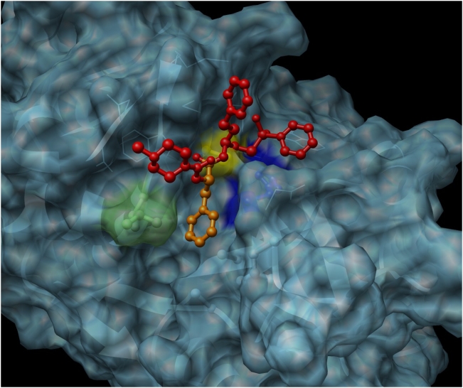
Figure 7. Structural model of SmCL3 in complex with the peptidyl vinyl sulfone inhibitor, K11777.
The inhibitor is shown in red and orange; the moiety that interacts with the S2 pocket is in orange. The catalytic Cys172and His317 residues are colored yellow and blue, respectively. The predicted residues in the deep S2 binding pocket are identical to those in human cathepsin V (used as a template for the model) except for a leucine residue (Leu216, colored light green).

