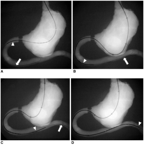
Fig. 3.
Radiographies of a coil catheter (arrowheads) being advanced over a guide wire (arrows) into the gastroduodenal phantom with stricture in the four groups.
A. Group I, without a balloon sheath.
B. Group II, through the deflated balloon sheath.
C. Group III, through the inflated balloon sheath with the inflated balloon located in the gastric fundus.
D. Group IV, through the inflated balloon sheath with the inflated balloon located in the gastric body.

