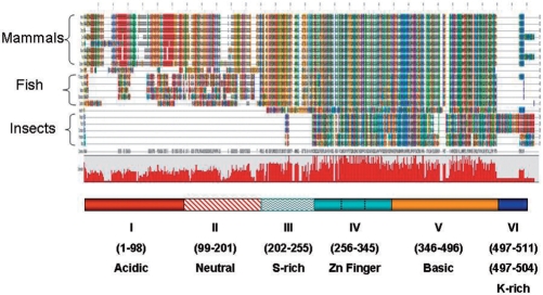
Figure 1.
Global protein sequence alignment of AEBP2 and JING. AEBP2 and JING protein sequences of different organisms were aligned using the ClustalW program. Different amino acids are represented in different colors and shades. The conservation level of each position is indicated in the graph below the alignment. Six conserved domains are indicated with different colors and patterns. The mouse AEBP2 protein was used as a reference to indicate the position of each conserved domain. The zinc finger and basic domains are the most conserved and show sequence conservation from flying insects to mammals. The zoom-in version of this alignment is available as Supplementary Data 5 or the following website (http://jookimlab.lsu.edu/?q=node/81).

