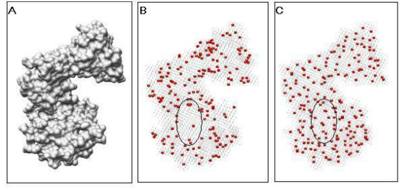
Figure 1.
Comparison of clustering sampling and random sampling techniques and effect. (A) The protein surface. (B) The red points depicted are the random sample points. (C) The red points depicted are the clustering sample points. Note that in figure(B) the ellipse marks a surface region where the sampled points distribute very sparsely, while the same surface region includes evenly distributed sampled points by using the clustering sample technique. The sample size is set at 300 in the figures.

