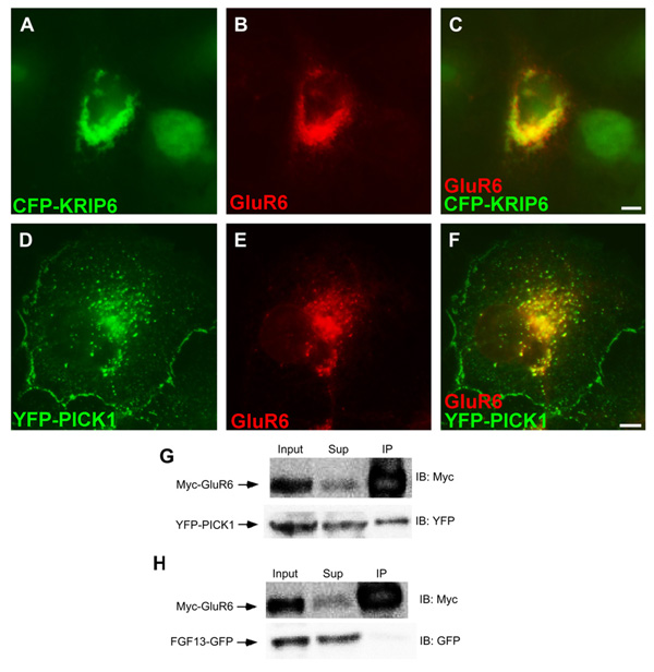
Fig. 2.
GluR6 co-clusters with KRIP6 and PICK1 and co-immunoprecipitates with PICK1. (A–F) Fluorescence images of COS-7 cells co-expressing GluR6, stained with a polyclonal anti-GluR6/7 antibody, visualized with an Alexa 568 (B, C) or Alexa 647 (E, F) conjugated secondary antibodies, and either CFP-KRIP6 (A–C) or YFP-PICK1 (D–F). CFP images and YFP images in A, C, D, F are shown in the green channel; GluR6 is shown in the red channel. Merged images (C, F) clearly indicate that GluR6 co-clusters with CFP-KRIP6 and with YFP-PICK1. Quantification of these interactions is illustrated in Fig. 3 panels I, J. Scale bars = 5 µm. (G) Immunoprecipitation with myc antibody resulted in co-immunoprecipitation of YFP-PICK1 from lysates of HEK cells stably expressing Myc-GluR6. Myc-GluR6 did not interact with FGF13-GFP. (H) The input and the supernatant collected after co-immunoprecipitation (Sup) represent 2.5% of the material used for immunoprecipitation. Following immunoprecipitation with anti-Myc-agarose beads (IP: Myc), immunoblots (IB) were performed with either the anti-YFP or the anti-Myc antibody (right).

