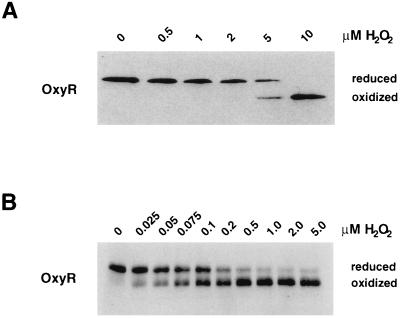
Figure 4.
Minimum concentrations of hydrogen peroxide required to oxidized OxyR. (A) Midlogarithmically growing wild-type cells (FÅ371) were treated with 0, 0.5, 1, 2, 5, and 10 μM hydrogen peroxide. (B) OxyR4C→A (0.01 μM final concentration) was reduced fully by incubation with a buffer containing 10 μM glutaredoxin 1, 25 mM GSH, and 0.1 mM GSSG. Aliquots were removed and treated with hydrogen peroxide to achieve 0, 0.025, 0.05, 0.075, 0.1, 0.2, 0.5, 1, 2, and 5 μM final concentrations. After 30 sec, the samples in both A and B were acidified with TCA and then treated with AMS. Again, separation and detection of OxyR was achieved by SDS/PAGE and immunoblot analysis.

