Fig. 1.
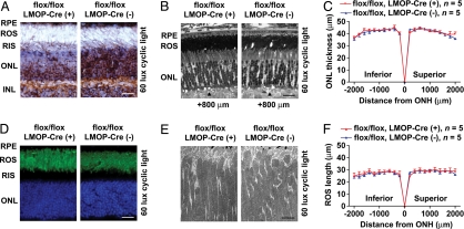
Photoreceptors of Rac1 CKO mice have normal structure. (A) Representative immunohistochemical staining of Rac1 (brown) in the retina from Rac1 CKO mice and WT litter-mates. Sections were stained with Mayer hematoxylin to show cell nuclei (light blue). (Scale bar: 20 μm.) (B) Morphology of the photoreceptor layer is normal in light microscopy of retinal sections cut along the vertical meridian containing the ONH. (Scale bar: 20 μm.) Arrowheads placed at 800 μm superior to ONH. (C and F) ONL thickness and ROS length of Rac1 CKO mice are indistinguishable from WT litter-mates at 20 points across the retina. (D) Representative immunohistochemical staining of rhodopsin (green) in the retina from Rac1 CKO mice and WT litter-mates. Nuclei in cells were stained with DAPI (blue). (Scale bar: 20 μm.) (E) Transmission electron micrographs showing the ROS structure of Rac1 CKO mice and WT litter-mates. (Scale bar: 2 μm.) RPE, retinal pigment epithelium; RIS, rod inner segments; INL, inner nuclear layer.

