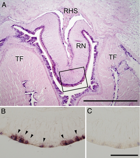
Fig. 5.
Localization of GSS mRNA in radial nerves by in situ hybridization. (A) The tissue around radial nerves (RN) was sectioned by using Technovit 7100 resin (Heraeus Kulzer GmbH, Wehrheim) and stained by hematoxylin-eosin to show the overall structure. TF, tube foot; RHS, radial hemal sinus. (Scale bar: 500 μm.) Isolated radial nerves were sectioned by using paraffin-embedding technique and were hybridized with DIG-labeled antisense (B) or sense (C) probes. Signals were detected in the peripheral region (arrows). The positions of B and C correspond to the square in A. (Scale bar: 100 μm.)

